Materials Needed:
- Skeletal models or specimens of the human skull
- Labels or markers
- Notebooks and pens
Procedure:
Introduction: The importance in protecting the brain, housing sensory organs, and providing attachment points for muscles.
Observation and Identification:
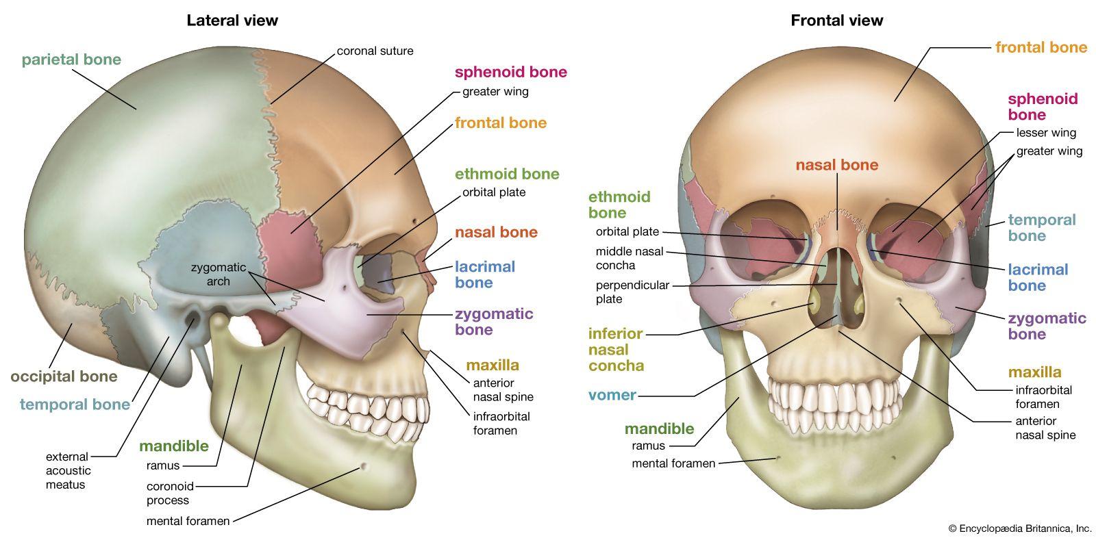
Carefully observe the skull and identify its key components, including:
Cranial bones: frontal, parietal (right and left), temporal (right and left), occipital, sphenoid, and ethmoid
Facial bones: maxilla (right and left), mandible, zygomatic (right and left), nasal (right and left), lacrimal (right and left), palatine (right and left), vomer, and inferior nasal conchae (right and left)
Note the shape, size, and relative positions of each bone.
Description: Once the bones are identified by the key features, describe each feature in detail. This can include:
Cranial bones:
- Frontal bone: frontal eminence, supraorbital margin, supraorbital foramen/notch
- Parietal bones: parietal eminence, sagittal suture, coronal suture, lambdoid suture
- Temporal bones: squamous part, tympanic part, mastoid process, styloid process, zygomatic process
- Occipital bone: foramen magnum, occipital condyles, external occipital protuberance
- Sphenoid bone: greater wings, lesser wings, sella turcica, pterygoid processes
- Ethmoid bone: cribriform plate, crista galli, perpendicular plate
Facial bones:
- Maxilla: alveolar process, infraorbital foramen, palatine process
- Mandible: body, ramus, angle, mental protuberance, mandibular foramen
- Zygomatic bones: zygomatic arch, zygomaticofacial foramen
- Nasal bones: bridge, nasal septum
- Lacrimal bones: lacrimal fossa
- Palatine bones: horizontal and perpendicular plates
- Vomer: nasal septum
- Inferior nasal conchae: nasal cavity
Discussion and Conclusion:
The importance of understanding the anatomy of the skull in fields such as anatomy, anthropology, and forensic science.
Safety Precautions: Handle skeletal models or specimens with care to avoid damage.
Leave a Reply